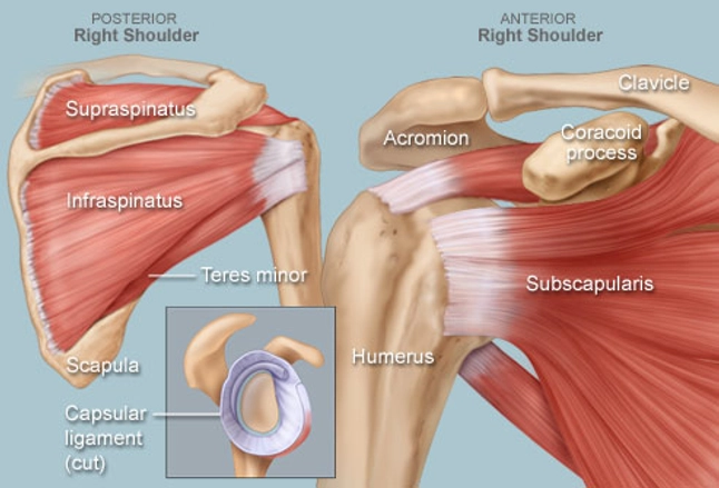
Shoulder Ligament Anatomy Diagram
Shoulder Ligament Anatomy Diagram. Glenohumeral, coracohumeral and transverse humeral ligaments movements: The clavicle (collarbone), the scapula (shoulder blade), and the humerus (upper arm bone) as well as associated muscles, ligaments and tendons. It is the major joint connecting the upper limb to the trunk. This diagram here just shows the joint capsule itself. The conoid and trapezoid ligaments make up the coracoclavicular ligaments. Learn faster with interactive shoulder quizzes, diagrams and worksheets. Although three ligaments protect and surround the shoulder joint, most. Robin smithuis and henk jan van der woude.
Additional stability is provided by: (1) the superior glenohumeral ligament (sghl), (2) the middle glenohumeral ligament (mghl), and (3) the inferior glenohumeral ligament (ighl). Normal anatomy, variants and checklist. 7 draw labelled diagram showing the relations of shoulder joint. Although the joint is held together by these extensive ligament and muscle attachments, certain types of forces can weaken the shoulder easily. Superior glenohumeral ligament and coracohumeral ligament are the primary restraints to posterior translation with the are flexed, adducted and internally acromioclavicular ligament anatomy. The human shoulder is made up of three bones: This page is about shoulder anatomy ligaments and muscles,contains soft tissues of the shoulder,shoulder joint; Human anatomy human body anatomy shoulder anatomy medical illustration human massage therapy ligament tear body anatomy for artists. All about the shoulder muscles.

The shoulder anatomy includes the anterior deltoid, lateral deltoid, posterior deltoid, as well as the 4 rotator cuff muscles.
The glenohumeral ligaments can be seen here, but they're not really. Muscle anatomy of the neck. This diagram here just shows the joint capsule itself. Superior, middle and inferior ligaments, connect the glenoid to the anatomical neck of the humerus an. Ac joint is a diathrodial joint with a fibrocartilaginous disk. There are several important ligaments in the shoulder. There are many shoulder ligaments which each play an important role in shoulder joint stabilization to various degrees: Arm flexion, extension, adduction, abduction the brachial plexus anatomy animation: Superior glenohumeral ligament and coracohumeral ligament are the primary restraints to posterior translation with the are flexed, adducted and internally acromioclavicular ligament anatomy. Static:gh ligaments, labrum & capsule and dynamic constraints: There are five major shoulder ligaments that keep the shoulder in place and prevent it from dislocating.
Glenohumeral joint,shoulder tendons,8 ejercicios para el hombro que debemos hacer and more. Although the joint is held together by these extensive ligament and muscle attachments, certain types of forces can weaken the shoulder easily. Webmd's shoulder anatomy page provides an image of the parts of the shoulder and describes its function, shoulder problems, and more. Although three ligaments protect and surround the shoulder joint, most of its stability comes from the powerful muscles and tendons of the rotator cuff. Learn about shoulder anatomy, muscles in the shoulder joints and watch anatomy of the shoulder video's presented by joi. An image depicting shoulder anatomy can be seen below. Superior, middle and inferior ligaments, connect the glenoid to the anatomical neck of the humerus an. There are several important ligaments in the shoulder. Robin smithuis and henk jan van der woude.
This diagram here just shows the joint capsule itself.
The primary function of the shoulder girdle is to give strength and range of motion to the arm. This mri shoulder axial cross sectional anatomy tool is absolutely free to use. Robin smithuis and henk jan van der woude. Rotator cuff & scapula stabilising. Notice superior labrum and attachment of the superior glenohumeral ligament. Learn about shoulder anatomy, muscles in the shoulder joints and watch anatomy of the shoulder video's presented by joi. There are five major shoulder ligaments that keep the shoulder in place and prevent it from dislocating. All about the shoulder muscles. The shoulder is not a single joint, but a complex arrangement of bones, ligaments, muscles, and tendons that is better called the shoulder girdle. 7 draw labelled diagram showing the relations of shoulder joint. Although three ligaments protect and surround the shoulder joint, most.
All about the shoulder muscles. Although the joint is held together by these extensive ligament and muscle attachments, certain types of forces can weaken the shoulder easily. Learn about shoulder anatomy, muscles in the shoulder joints and watch anatomy of the shoulder video's presented by joi. There are several important ligaments in the shoulder.
Roots, trunks, divisions, cords, branches, clinical anatomy.
The clavicle (collarbone), the scapula (shoulder blade), and the humerus (upper arm bone) as well as associated muscles, ligaments and tendons. Glenohumeral joint,shoulder tendons,8 ejercicios para el hombro que debemos hacer and more. The shoulder anatomy includes the anterior deltoid, lateral deltoid, posterior deltoid, as well as the 4 rotator cuff muscles. A joint capsule is a watertight sac that surrounds a joint. Muscle anatomy of the neck. Bones in shoulder, ligaments of the shoulder joint, parts of the shoulder joint, shoulder anatomy, shoulder joints and muscles, shoulder structure anatomy, shoulder tendon anatomy, shoulder related posts of diagram of shoulder muscles and tendons. Static:gh ligaments, labrum & capsule and dynamic constraints: It is the major joint connecting the upper limb to the trunk. The primary function of the shoulder girdle is to give strength and range of motion to the arm. Glenohumeral, coracohumeral and transverse humeral ligaments movements:
Ligaments are soft tissue structures that connect bones to bones shoulder anatomy diagram. You can see it enclosing the glenohumeral joint and you can see its attachment on the anatomical you've got the transverse humeral ligament and the coracohumeral ligament.

Posting Komentar untuk "Shoulder Ligament Anatomy Diagram"